
Series description
Pectus excavatum is a natural deformity to the chest, which causes it to be concave. It can cause a lot of health complications and in my case it not only affected my physical endurance, but my self-image as well. This led me to one of the most life-defining experiences I have had: a surgery called the Nuss procedure.
The Nuss procedure is regarded as minimally invasive, but it is one of the most painful surgeries a child can go through. This procedure has often been likened to that of braces, but instead of taking years to work, it happens in just a few seconds. Although there were four tough years of pain while I recovered, I would go through it all again if I had to. This project focuses on the pain and clinical nature of surgery, as well as the beauty of the human body.
Biography
Austin Crail is a freelance photographer working in Philadelphia, PA. He grew up in rural New Jersey where he learned to have a strong sense of community. While his time living in Shanghai gave him a deep respect for all. Graduating with a BS in photography from Drexel University taught him all he needs to know about commercial and fine art photography. Even so, he is always eager to refine his craft and strengthen his style.
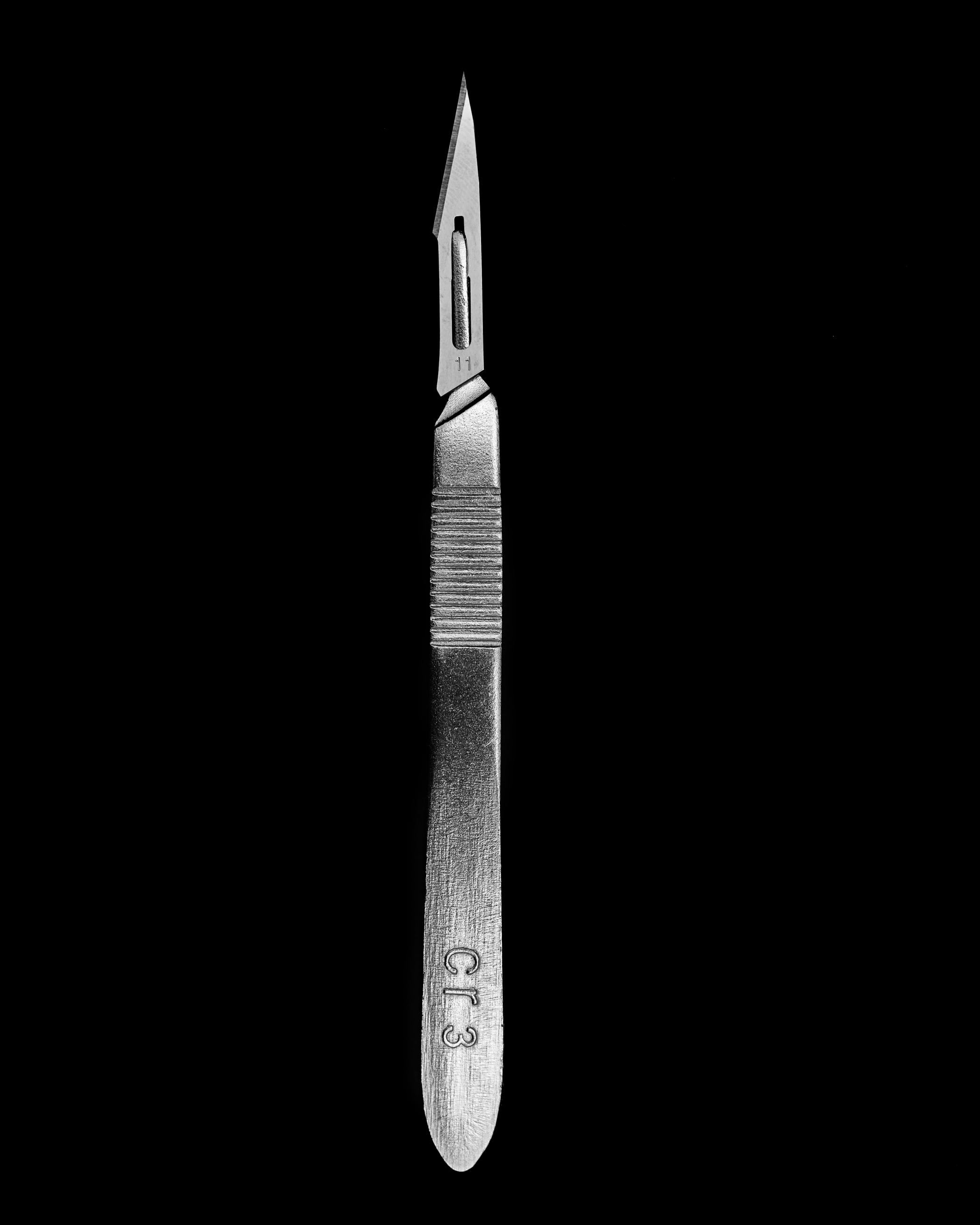
Scalpel
I took this in a studio with a big softbox looming over the scalpel, which was sitting on a black background. I wanted to show the similarities not only between the shape of the scar and scalpel, but the texture as well.
© Austin Crail, United States, Shortlist, Professional competition, Still Life, 2023 Sony World Photography Awards
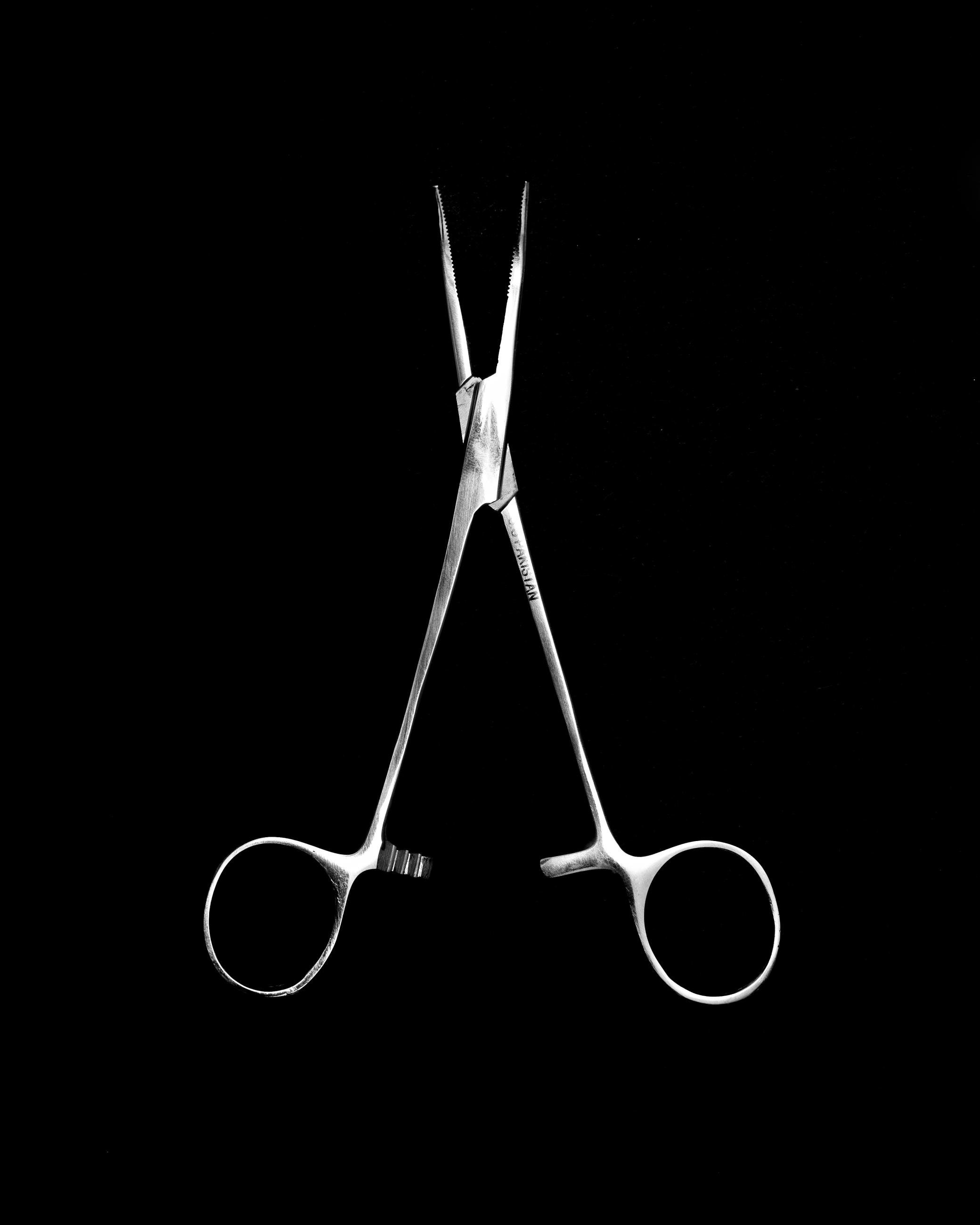
Forceps
Forceps are used to align and flip the steel bar that is inserted into the chest during the Nuss procedure, instantly popping out and reshaping the ribcage. Shot on a black background with a large softbox over the subject, I wanted to mimic the way a patient would feel on the operating table.
© Austin Crail, United States, Shortlist, Professional competition, Still Life, 2023 Sony World Photography Awards
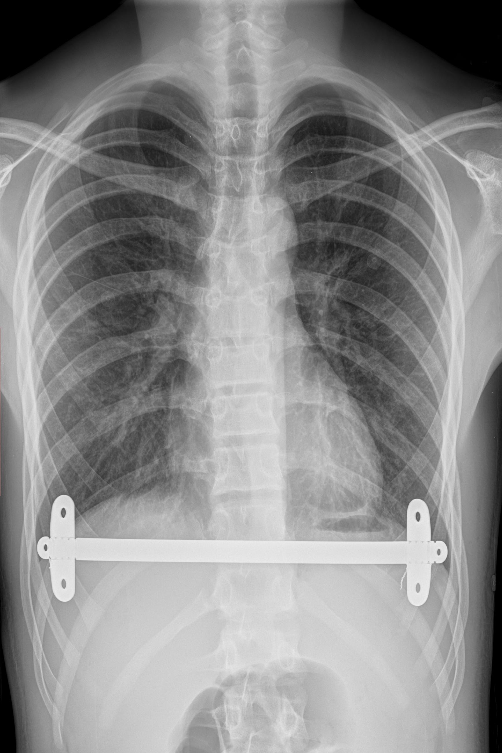
X-ray
An X-ray of the steel bar inside my body, under my rib cage and over my heart and lungs. The image was modified slightly to remove some text over the image and to show more of the human form. This image and the photograph of the front of my chest form a diptych, with each image echoing the other.
© Austin Crail, United States, Shortlist, Professional competition, Still Life, 2023 Sony World Photography Awards

Scar
This scar shot is made to mimic the photograph of the scalpel. It was lit with a single light, carefully placed so the hair would catch the light and also to reveal the texture of the skin. It shows the depth of the scar to contrast the ‘minimally invasive’ nature of the surgery.
© Austin Crail, United States, Shortlist, Professional competition, Still Life, 2023 Sony World Photography Awards
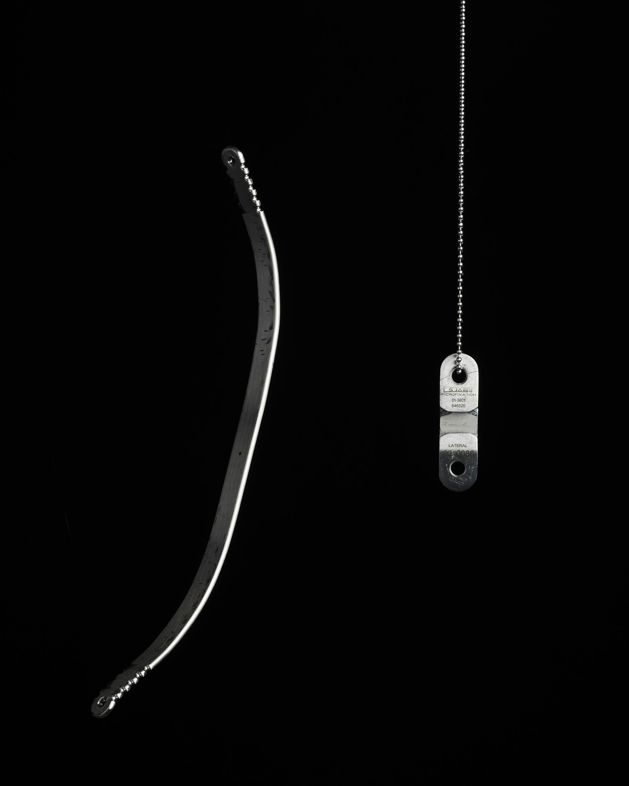
Bar and Stabilizer
This is the bar that was placed over my heart and under my ribs, along with one of two stabilisers that were screwed into my ribcage. The stabilisers were located either side of the chest and held the bar in place for four years.
© Austin Crail, United States, Shortlist, Professional competition, Still Life, 2023 Sony World Photography Awards
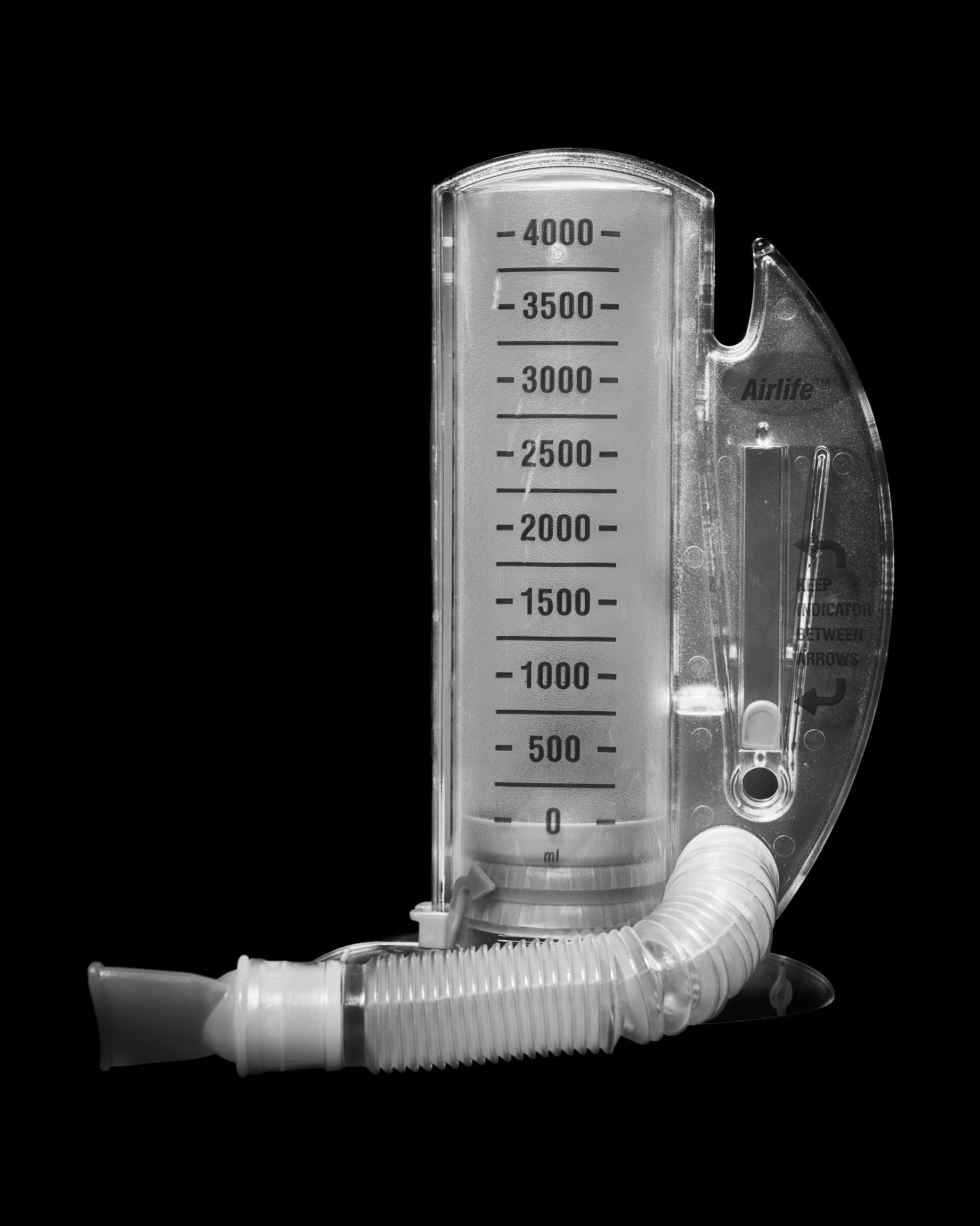
Lung Strength Test
I photographed this because it was not only used to test my lung strength after my operation, but it was also a crucial test in determining whether or not my medical insurance would help cover the operation in the first place. The insurers needed to determine whether or not it was purely a cosmetic surgery, or if the condition was impacting me physically.
© Austin Crail, United States, Shortlist, Professional competition, Still Life, 2023 Sony World Photography Awards
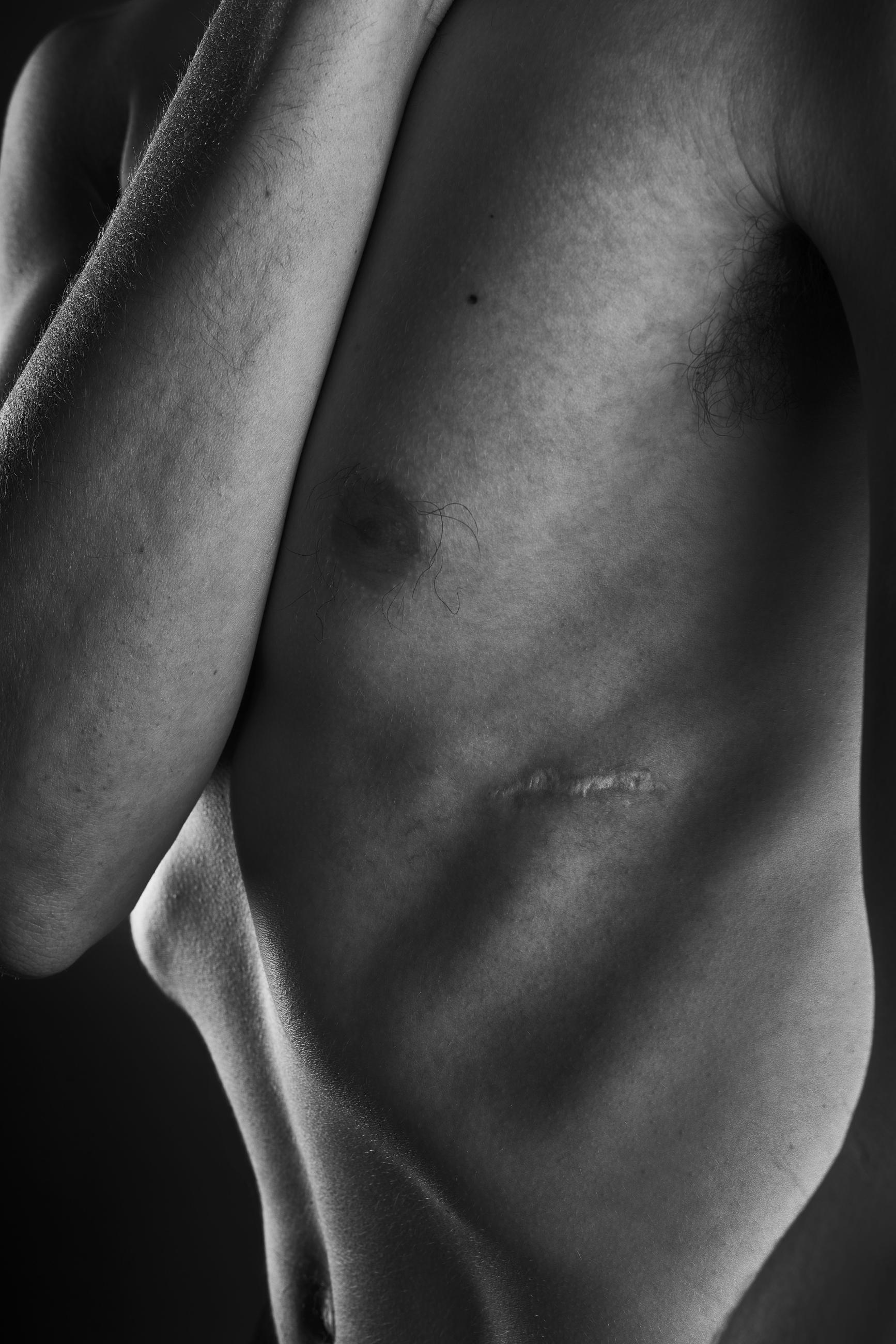
Scar Left
I took this photograph with the arms up to highlight the vulnerability of humans, especially those affected by pectus excavatum. I used a two light setup; one light to graze the scar and emphasise the ridges of the ribcage, and the other to draw attention to the form of the body itself.
© Austin Crail, United States, Shortlist, Professional competition, Still Life, 2023 Sony World Photography Awards